

Somatic Dentistry
Reading the evidence on teeth and the brain
A research archive that reads the original studies linking the teeth, brain and body, and keeps established anatomy apart from connections that are still hypotheses.
- Established (solid)
- Observed in human studies (dashed)
- Still a hypothesis (dotted)
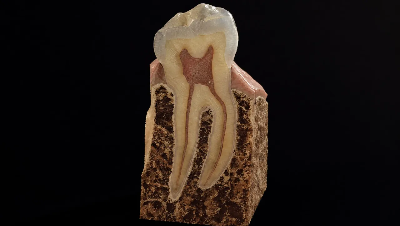
- 1Station 1: Occlusion Concept visualization Link to the next station: established (solid line)
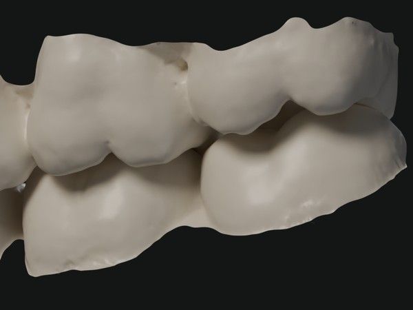
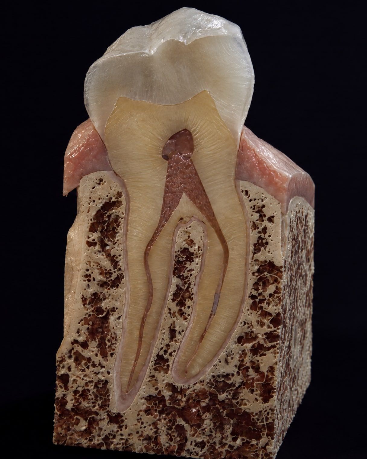
- 2Station 2: Periodontal ligament Concept visualization · redrawn with AI tools Link to the next station: established (solid line)
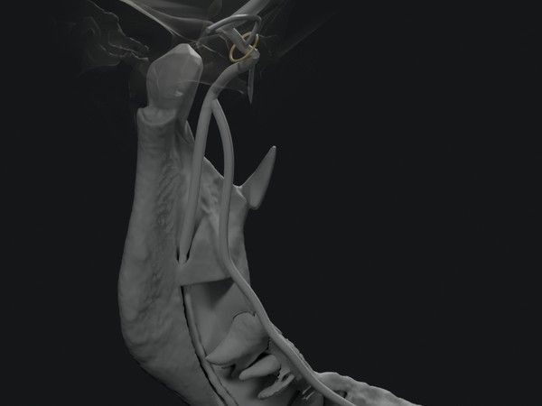
- 3Station 3: Trigeminal nerve Concept visualization Link to the next station: established (solid line)
- 4Station 4: Brainstem Concept visualization Link to the next station: still a hypothesis (dotted line)
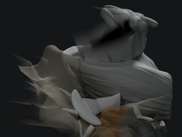
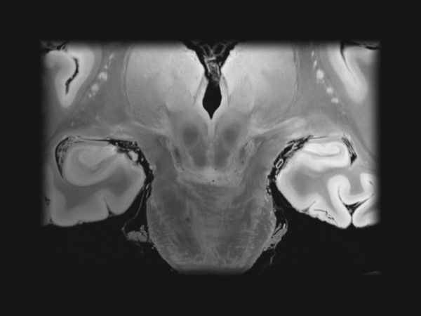
- 5Station 5: Hippocampus Real image · 7T MRI Link to the next station: not yet determined
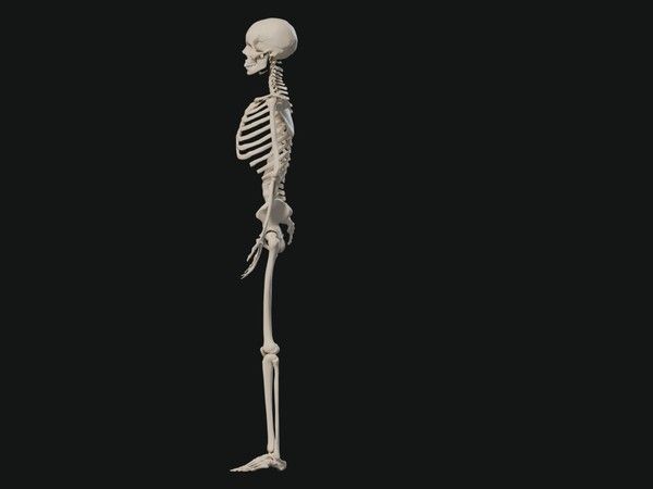
- 6Station 6: Whole body Concept visualization
In English
- Definition
What is Somatic Dentistry?
- Reading the evidence
How small a force can a tooth feel?
The sense behind holding half a peanut with less than 1 N
More English pages are in preparation. The Korean site has more pages; use the KO / EN switch at the top of any page.
Definition
What is Somatic Dentistry?
Somatic Dentistry is a perspective within dentistry that reads how sensation and movement in the teeth and mouth connect with the rest of the body, distinguishing connections established by anatomy, those observed in human studies and those that are still hypotheses.
Anatomical journey
From occlusion to the brain,
six stations.
Each station has a name, a one-line note and its level of evidence. Established stretches and those still open to question are told apart on the same screen.
Station 01
Occlusion
How the upper and lower teeth come together
Station 02
Periodontal ligament (PDL)
Mechanoreceptors in the PDL around the root pick up the force placed on the tooth. Each tooth has several hundred of these receptors.
EstablishedStation 03
Trigeminal nerve
Periodontal signals travel through the alveolar nerves to the trigeminal ganglion, and these signals have also been recorded from the inferior alveolar nerve in people.
EstablishedStation 04
Brainstem
Signals enter the trigeminal sensory nuclei of the pons and medulla, and the brainstem’s chewing rhythm generator uses this information to choose the chewing movement.
EstablishedStation 05
Hippocampus
The idea that sensory input from the teeth is linked to memory and cognition.
Still a hypothesisStation 06
Whole body
- ⑤ Hippocampus → ⑥ Whole bodyNot yet determined
- ① Occlusion → ⑥ Whole-body postureObserved in human studiesFindings are mixed
The relationship between occlusion and body posture. Some studies have reported a relationship and others have not; where correlations are reported they are weak, and neither a predictable relationship nor causation has been confirmed.