

Insight · Reading the evidence
How small a force can a tooth feel?
The sense behind holding half a peanut with less than 1 N
Teeth feel forces far smaller than 1 N (about the weight of a tangerine) through mechanoreceptors in the periodontal ligament (PDL) around their roots1,2.
For most of these receptors, the range of greatest sensitivity is 1 N or less at the front teeth and 4 N or less at the back teeth, so it is smaller toward the front3.
The force needed for a person to notice a tooth being pressed is far smaller still: a periodontology textbook gives 1–2 g (about 0.01–0.02 N)1, and one study found a mean of about 0.11 N2.
The force the front teeth use to hold half a peanut (under 1 N) also falls within the receptors’ most sensitive range4,5, and the researchers concluded that this sense is used mainly when food is held and handled lightly5.
The idea that this sense extends as far as memory or cognition, however, is still a hypothesis.
Contents of this article
Tracing tooth sensation with half a peanut
How much force do the front teeth use to hold a peanut?
The front teeth hold half a peanut with a force of less than 1 N4,5. When a Swedish research group asked people to hold half a peanut between their central incisors and then split it, splitting took 16–19 N, more than twenty times the holding force4. One newton (1 N) is roughly the weight of a tangerine resting in your palm. Teeth feel more than pain. When we bite, they also feel how much force is on them, and that sense reaches down to very small forces. The jaw muscles produce the force, but sensation at the tooth root also helps to gauge forces this small5,6.
Table 1 Forces when holding and splitting with the incisors. The newton (N) is the unit of force; 1 N is about the weight of a 100 g object (site calculation). A tangerine weighs 76–120 g (USDA FoodData Central, tangerine FDC 169105).
| Phase | Force | Also observed |
|---|---|---|
| Holding4 | Under 1 N; 0.36–0.76 N across participants (biscuit and peanut alike) | Steady during each hold and similar from one attempt to the next |
| Splitting4 | Peanut: 16.0–19.0 N across participants | More than twenty times the holding force (site calculation) |
Where does a tooth feel such a small force?
A tooth feels such small forces mainly through sensory receptors in the periodontal ligament (PDL) that surrounds its root1,6. A tooth is not attached directly to the bone. Between the root and the bone lies a gap a few hairs wide (0.15–0.38 mm)7,8. In each tooth, the PDL that fills this gap holds several hundred mechanoreceptors, which turn force into nerve signals6.
Even a single incisor has several hundred of these sensory endings6, and most of them respond most keenly to changes in force below 1 N, less than the weight of a tangerine3. That is precisely the level of force the incisors used to hold the peanut.
A gap a few hairs wide holds this much sensation. That is why I see the inside of the mouth not as a small space, but as one as vast and intricate as the universe. That space is built a little differently at each tooth: toward the molars, the force used to hold a peanut and the range in which most receptors are sensitive grow larger together3,9.
The force at which a person first notices a tooth being pressed is far smaller still: the periodontology textbook gives 1–2 g1. The value varies from study to study and from person to person2, but on average it stays far below the force used to hold a peanut.
Read the graph as text
| Tooth | Front or back | Force used to hold half a peanut (20 people) | Range where most receptors are most sensitive |
|---|---|---|---|
| Central incisor | Front teeth | 0.60 N | 1 N or less |
| Canine | Front teeth | 0.77 N | 1 N or less |
| Second premolar | Back teeth | 1.15 N | 4 N or less |
| First molar | Back teeth | 1.74 N | 4 N or less |
| Natural teeth (tooth type not specified) | Not specified | Force first felt: 1–2 g in the textbook (about 0.01–0.02 N, passive force detection); mean about 0.11 N in one study (22 people; range 1.2–26.2 g across participants) | Not applicable |
The canine is grouped with the front teeth; its sensitive range was not measured separately. The sensitive range starts at 0 N: for the front teeth the text of the review (Trulsson 2006) gives 0–1 N, and for the back teeth the review gives only an upper limit of 4 N. The sensitive range comes from nerve recordings and the force first felt from people's responses, so they are different measurements. The force first felt varies by study and by person; most of the textbook range (1.2–2 g of its 1–2 g) lies within one study's range across participants (1.2–26.2 g). Holding force, sensitive range and force first felt come from different publications; putting them together is the site's choice.
How does tooth sensation become jaw-muscle force?
Sensory signals from the teeth travel along the trigeminal nerve to the brainstem10 and are used to regulate the force of the jaw muscles5. An occlusion textbook explains that the brainstem’s chewing rhythm generator (a central pattern generator) uses this information to choose the most suitable chewing movement10. According to the pathway mapped in animal studies, the signal then passes to the motor neurons of the jaw-closing muscles11. When the muscles change their force, that change reaches the PDL again, so in effect the signal travels a full loop. This loop is the site’s summary, weaving together textbook explanations and a number of experiments.
A touch of less than 1 N at the tip of an incisor travels along the trigeminal nerve to the brainstem and comes back as force in the jaw-closing muscles. This loop is the site’s summary, and the details of the last stretch, from the brainstem to the motor neurons, rest on animal studies.
The teeth seem to be one thing and the jaw muscles another. Yet even while holding just half a peanut, the two do not move separately. I read this loop as one of the smallest scenes showing that the body is connected.
The whole loop. Pick a step to light up only that part.
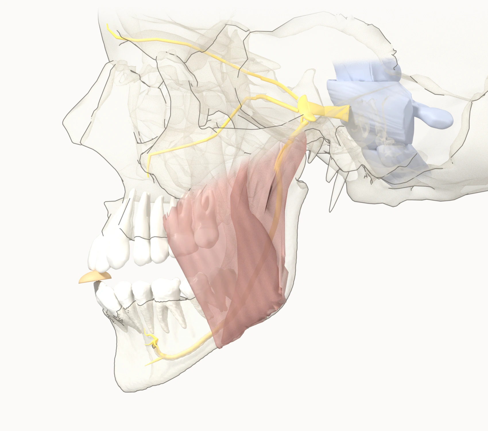
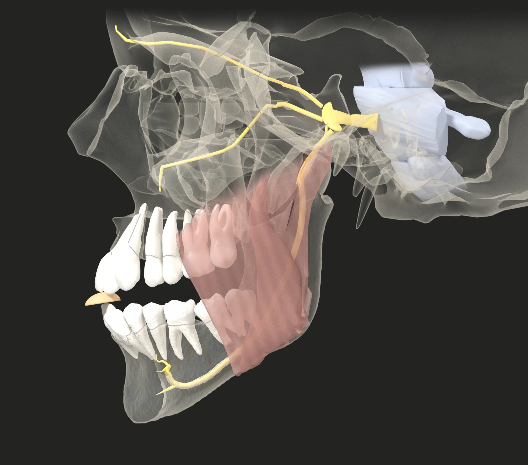

- Textbook anatomy and human experiments (some details from animal data)
- Experiments that reduced periodontal sensation
- Still a hypothesis
- Gray: other sensory input (muscle spindles)
Anatomy: lower jaw, teeth and inferior alveolar canal from ToothFairy2 CBCT (Bolelli et al.) — the upper ramus (condyle, coronoid process) completed from Z-Anatomy · skull (including the facial bones and upper jaw), upper teeth, brainstem, nerves and masseter from Z-Anatomy (Gauthier Kervyn, Marcin Zielinski; based on BodyParts3D) · root entry position from 7T MRI (Edlow et al., CC0) · root direction from HCP1065 (Yeh 2022, Human Connectome Project data). Lower jaw (public CBCT data, one person) and skull with upper jaw (anatomical atlas model) are from two different people, aligned as stated under “Modifications” in the credits. The peanut-holding pose is computed. The positions of the trigeminal ganglion, nerve roots and trigeminal nuclei are schematic, as are the short connections from the ganglion to each opening. Adapted from the original data. This figure is licensed under CC BY-SA 4.0.
Full credits
- ToothFairy2 dataset (University of Modena and Reggio Emilia · Radboud University), CC BY-SA 4.0. Case ToothFairy2P_091, labels 1 (mandible), 3 (left inferior alveolar canal) and 31–38 (left lower teeth) only. Bolelli F et al., Med Image Anal 2026;104095, https://doi.org/10.1016/j.media.2026.104095 · Bolelli F et al., CVPR 2025, https://doi.org/10.1109/CVPR52734.2025.00494 · Lumetti L et al., IEEE Access 2024, https://doi.org/10.1109/ACCESS.2024.3408629
- Z-Anatomy (Gauthier Kervyn, Marcin Zielinski), CC BY-SA 4.0: skull (skull base, frontal, parietal, nasal, lacrimal, ethmoid, maxilla, palatine, vomer, inferior nasal conchae, zygomatic), upper teeth (11–17, 21–27), brainstem, nerve curves, masseter, completion of the upper ramus. Many shapes are based on BodyParts3D, © The Database Center for Life Science, CC BY 4.0.
- Edlow BL, Mareyam A, Horn A, et al. 7 Tesla MRI of the ex vivo human brain at 100 micron resolution. OpenNeuro ds002179, https://doi.org/10.18112/openneuro.ds002179.v1.1.0, CC0.
- Tractography: HCP1065 atlas, Yeh FC (2022), CC BY-SA 4.0, https://doi.org/10.1038/s41467-022-32595-4. Data were provided in part by the Human Connectome Project, WU-Minn Consortium (Principal Investigators: David Van Essen and Kamil Ugurbil; 1U54MH091657) funded by the 16 NIH Institutes and Centers that support the NIH Blueprint for Neuroscience Research; and by the McDonnell Center for Systems Neuroscience at Washington University. · WU-Minn HCP Consortium Open Access Data Use Terms
- Modifications: rendered with a new camera, translucency and outlines, the top of the skull faded; the lower jaw rotated and shifted to sit closed against the skull (the mismatch between the two datasets was shared among connected structures, such as the condyle's position in the jaw joint and the relation of the upper and lower teeth, according to how much each can move or adapt; this is a fitted relation, not one person's anatomy); as part of that fit, the upper jaw with its teeth moved back by 3.9 mm and the upper incisors and canines tilted by 4° (the maxilla's shift tapers to zero below the orbit and also moves the distal part of the maxillary nerve that runs through the upper jaw); the peanut-holding pose is computed (the lower jaw protruded, with the condyle about 8 mm forward, and opened by about 5°, computed for this model's bone shape); the right half of the lower jaw, teeth 41–48 and the right temporal bone are not drawn (hidden in the side view); the insertion of the (atlas) masseter moved to that jaw position and vertices inside the bone pushed out by up to 3.4 mm; arrows, text and the peanut added.
- License: CC BY-SA 4.0, https://creativecommons.org/licenses/by-sa/4.0/
Read the figure as text
- ① Contact and sensing When the peanut touches the incisors, the mechanoreceptors in the PDL respond. At the incisors, most of them are most sensitive to force changes of 1 N or less.
- ② From brainstem to jaw muscle The signal travels along the trigeminal nerve to the brainstem and feeds into the activity of the jaw-closing muscles. Other sensory input, such as from muscle spindles, comes in as well. The detailed wiring from the brainstem to the motor neurons rests on animal data.
- ③ From holding to splitting First the peanut is held with under 1 N, then split with 16–19 N. While it is held, the PDL senses the force changes again.
- ④ With PDL sensation reduced When the periodontal tissues were anesthetized, the holding force grew larger and, in some experiments, unsteady. The size of the splitting force stayed the same, but one study found that the time taken to split the peanut and the rate at which force rose both changed.
Links in the figure
- Peanut contact → PDL
- PDL → Trigeminal nerve (signal)
- Trigeminal nerve → Brainstem
- Brainstem → Jaw-closing muscle (regulates activity)
- Jaw-closing muscle → Hold
- Hold → Split
- Hold → PDL (force change sensed again)
- Muscle spindles etc. → Trigeminal nerve
- PDL anesthesia → Trigeminal nerve (reduced signal, blue line)
- PDL anesthesia → After anesthesia (blue line)
- PDL → Wider brain functions (still a hypothesis, dotted line)
What changes when the periodontal tissues are anesthetized?
When the periodontal tissues are anesthetized, the force used to hold a peanut grows larger, and the morsel often slips from the incisor tips4,5. Some experiments found that the force also became unsteady, but in the largest one (45 people) it did not become more unsteady4,5,9,12. Anyone who has tried to pick up a small bead while wearing thick gloves will recognize the picture. The size of the splitting force stayed the same4,5,9, but in one study (15 people) splitting the peanut took longer and the force rose more slowly13.
When the sensation was briefly reduced, holding became clumsy, but the large splitting force stayed the same. The researchers concluded that this sense is used mainly when food is held and handled lightly, rather than when it is bitten hard5.
Read the graph as text
| Condition | Holding force | Splitting force (peanut) |
|---|---|---|
| Normal sensation | Under 1 N; 0.36–0.76 N across participants | 16.0–19.0 N across participants (more than twenty times the holding force; site calculation) |
| Periodontal sensation reduced (local anesthesia) | Larger and unsteady (no value in the abstract) | No effect |
Data: Trulsson & Johansson 1996b. The arrow and the wave mark for the holding force after anesthesia show direction only: the abstract gives no value, and the same group's review (Trulsson & Johansson 1996a) shows the values only in a figure.
Table 2 What changed when periodontal sensation was reduced (local anesthesia)
| Experiment (participants) | What changed |
|---|---|
| Holding and splitting a morsel with the incisors4,5 | Holding force larger and unsteady. The morsel often slipped from the incisor tips. No effect on the splitting phase |
| Half a peanut, incisors to molars (4 people)9 | Holding force larger and more unsteady at every tooth. No clear change in the splitting phase |
| Biscuit and peanut (15 people)13 | Before anesthesia biscuits split at about 9 N and peanuts at about 18 N on average; after anesthesia, splitting a peanut took longer and the force rose more slowly, so the force profiles for the two foods could no longer be told apart |
Show the other experiments
| Experiment (participants) | What changed |
|---|---|
| Chewing (14 people; infiltration and PDL injections on the chewing side)14 | Number of chewing strokes unchanged; timing of masseter (a chewing muscle) activity changed. The researchers estimate that periodontal sensation contributes about 20% of the muscle activity in the jaw-closing phase |
| 45 people in three anesthesia groups (reference group: 20 people from an earlier study with the same task)12 | Holding force higher in the incisor and nerve-block groups. Holding and splitting forces did not become more unsteady, and splitting force did not differ between groups. With repetition, force tended to recalibrate |
| Another research group, holding a light sensor between the incisors (20 people)15 | When seated, the minimum force needed to keep the sensor from dropping rose from a mean of 0.63 N to 1.87 N |
| Another research group, periodontal tissues and mucosa anesthetized on the chewing side (11 people)16 | Reduced ability to break up and mix peanuts |
To return to the half peanut we began with: that light force of less than 1 N fell within the range in which most of the sensory receptors at the tooth root are most sensitive, and when that sensation was reduced, the change centered on that same light hold. In the next article, I plan to read the evidence on how this loop changes with implants, which have no PDL.
The mouth is not the only window into the body. Still, I think it is one of the high-resolution ones: a window that can feel even forces far smaller than 1 N.
This note covers the PDL, the second station on the Somatic Dentistry evidence map. Definition: Somatic Dentistry is a perspective within dentistry that reads how sensation and movement in the teeth and mouth connect with the rest of the body, distinguishing connections established by anatomy, those observed in human studies and those that are still hypotheses. What is Somatic Dentistry? · Evidence map: PDL (coming soon)
Evidence
- What this article can say. When people hold food with their incisors, they tend to use the small range of forces to which periodontal mechanoreceptors are most sensitive, and reducing periodontal sensation makes the holding force larger.
- What this article cannot say. It does not show that the PDL alone sets the force, that the anesthesia results reflect the effect of the PDL alone, or that training this sense improves health or brain function.
The table below collects, for each of the four questions in the text, the supporting evidence alongside contrary results and limitations; evidence beyond those questions (the FAQ and the analogies) is in the last row. Each cell starts with the broadest sources: textbooks, then reviews and systematic reviews, human studies, animal studies and hypotheses. Textbooks are ordered by how directly they state the row’s claim rather than by citation count; other sources in the same tier are ordered by citation count (OpenAlex, retrieved October 11, 2026). In the supporting-evidence column, each cell shows representative sources, and the rest can be expanded. In the contrary-results column, textbooks come first, and the rest can be expanded. “Same research line” marks publications with Trulsson as an author. There are five grades: established (textbook), human studies, animal data, hypothesis and editorial interpretation.
Table 3 The four questions in the text and further evidence
| Question in the text | Supporting evidence | Contrary results and limitations | Grade |
|---|---|---|---|
| How much force do the front teeth use to hold a peanut? Under 1 N; splitting takes more than twenty times that |
3 more (3 human studies)
|
2 more contrary results and limitations
| Human studies |
| Where does a tooth feel such a small force? Mainly by the PDL mechanoreceptors around the root. The receptors’ most sensitive range (nerve recordings) and the force a person first feels (people’s responses) are different measurements |
4 more (2 textbooks, 1 review, 1 human study)
|
1 more limitation
| Established, human studies |
| How does tooth sensation become jaw-muscle force? It travels along the trigeminal nerve to the brainstem and regulates jaw-muscle force. The whole loop (Figure 1) is the site’s summary |
8 more (3 reviews or systematic reviews, 2 human studies, 2 animal studies, 1 hypothesis)
|
1 more limitation
| Established, human studies, animal data (brainstem details), hypothesis (dotted line in Figure 1) |
| What changes when the periodontal tissues are anesthetized? The holding force rises. Some experiments found it became unsteady and the largest (45 people) did not; the splitting force kept its size, though one study found splitting took longer |
9 more (1 textbook, 1 review, 7 human studies)
|
2 more contrary results and limitations
| Established, human studies |
| Further evidence (FAQ, analogies, conversions) Implant sensation, brain function, and analogies that give a sense of scale |
5 more (3 human studies, 1 hypothesis, 1 analogy)
|
| Established, human studies, hypothesis, editorial interpretation |
Show the evidence for every statement
Table 4 Claims table: the evidence grade and sources of the 32 main factual statements in the text (numbers refer to the reference list)
| Claim | Evidence grade | Source no. · DOI |
|---|---|---|
| Periodontal touch information is used mainly for the fine control of biting and chewing forces, not for protection | Established | 1 ISBN 978-0-470-67248-8 |
| Teeth feel the forces placed on them as touch (periodontal receptors provide the conscious perception of force) | Established | 1 ISBN 978-0-470-67248-8; 2 doi:10.1034/j.1600-0501.1995.060203.x; 6 doi:10.1016/b978-0-7234-3809-0.00010-3 |
| The force at which a pressed tooth is first felt is 1–2 g (about 0.01–0.02 N) in the textbook, and 50–100 g for implants | Established | 1 ISBN 978-0-470-67248-8 |
| A loop from tooth sensation through the trigeminal nerve, brainstem and jaw muscles back to the teeth (Figure 1; the site's summary of textbooks and studies) | Editorial interpretation | 1 ISBN 978-0-470-67248-8; 4 doi:10.1007/BF00230428; 5 doi:10.1016/s0301-0082(96)00016-0; 10 ISBN 978-0-323-58210-0; 11 doi:10.2334/josnusd.16-0845; 26 doi:10.1177/0022034509352842; 27 doi:10.1007/BF00247900 |
| Toward the molars the holding force and the receptors' sensitive range grow together (two results placed side by side) | Editorial interpretation | 1 ISBN 978-0-470-67248-8; 2 doi:10.1034/j.1600-0501.1995.060203.x; 3 doi:10.1111/j.1365-2842.2006.01629.x; 9 doi:10.1007/s00221-006-0719-9 |
| The receptors' most sensitive range (nerve recordings) and the force a person first feels (people's responses) are different measurements, and the force first felt varies by study and person | Editorial interpretation | 1 ISBN 978-0-470-67248-8; 2 doi:10.1034/j.1600-0501.1995.060203.x; 3 doi:10.1111/j.1365-2842.2006.01629.x; 4 doi:10.1007/BF00230428 |
| When a natural tooth was pressed with steadily increasing force, the force at which this was first felt averaged 11.5 g (about 0.11 N); implants needed 8.75 times that (22 people) | Human studies | 2 doi:10.1034/j.1600-0501.1995.060203.x |
| Most periodontal mechanoreceptors are most sensitive to force changes of 1 N or less at the front teeth and 4 N or less at the back teeth | Human studies | 3 doi:10.1111/j.1365-2842.2006.01629.x |
| The force for holding half a peanut or a piece of biscuit with the central incisors is under 1 N (0.36–0.76 N across participants) and steady | Human studies | 4 doi:10.1007/BF00230428 |
| The force for splitting half a peanut is 16.0–19.0 N across participants | Human studies | 4 doi:10.1007/BF00230428 |
| With the periodontal tissues anesthetized, the holding force is larger and unsteady and the morsel slips out, while the splitting phase is unaffected | Human studies | 4 doi:10.1007/BF00230428; 5 doi:10.1016/s0301-0082(96)00016-0 |
| 1 N is about the weight of a 100 g object, and the splitting force is more than twenty times the holding force (site calculation) | Editorial interpretation | 4 doi:10.1007/BF00230428 |
| 1 N is about the weight of a tangerine resting in the palm (one tangerine 76–120 g; analogy) | Editorial interpretation | 4 doi:10.1007/BF00230428; fdc.nal.usda.gov |
| The anesthesia result is compared with picking up a small bead in thick gloves (analogy) | Editorial interpretation | 4 doi:10.1007/BF00230428; 5 doi:10.1016/s0301-0082(96)00016-0; 9 doi:10.1007/s00221-006-0719-9 |
| Human periodontal signals were recorded from the inferior alveolar nerve, which supplies the lower teeth. The researchers concluded that this sense is used mainly when food is first contacted, handled and held | Human studies | 5 doi:10.1016/s0301-0082(96)00016-0 |
| Each tooth's PDL holds several hundred highly sensitive touch receptors; none lie close to an implant | Established | 6 doi:10.1016/b978-0-7234-3809-0.00010-3 |
| Whether the jaw-opening reflex is needed for chewing or serves only a protective role is not known | Established | 7 ISBN 978-0-323-48524-1 |
| The PDL is soft connective tissue between root and bone, 0.15–0.38 mm wide | Established | 7 ISBN 978-0-323-48524-1 |
| The PDL is as wide as a few hairs side by side (one hair about 80 µm; analogy) | Editorial interpretation | 7 ISBN 978-0-323-48524-1; 8 doi:10.1155/2020/2476890 |
| The force for holding half a peanut rises from 0.60 N at the central incisor to 1.74 N at the first molar; with anesthesia it rises and becomes unsteady at every tooth | Human studies | 9 doi:10.1007/s00221-006-0719-9 |
| The brainstem's chewing rhythm generator chooses the chewing movement from the information the teeth and PDL feed back | Established | 10 ISBN 978-0-323-58210-0 |
| Sensory signals from the teeth and mouth enter the brainstem (pons) along the trigeminal nerve | Established | 10 ISBN 978-0-323-58210-0 |
| The mesencephalic trigeminal nucleus in the brainstem receives input from the PDL and muscle spindles and relays it mainly to the jaw-closing motor neurons | Animal data | 11 doi:10.2334/josnusd.16-0845 |
| With anesthesia, splitting a peanut takes longer and the force rises more slowly (before anesthesia, biscuits split at about 9 N and peanuts at about 18 N) | Human studies | 13 doi:10.1111/j.1600-0722.2009.00691.x |
| Implants also give a sense of touch, known as osseoperception (systematic review of human and animal studies) | Human studies | 22 doi:10.1111/joor.13296 |
| With a single implant opposed by a natural tooth, detecting a thin foil was about as good as between natural teeth (62 people) | Human studies | 23 doi:10.1111/j.1600-0501.2006.01321.x |
| In mammals, a pattern generator in the brainstem (pons and medulla) generates the chewing rhythm, and feedback from oral touch receptors, muscle spindles and PDL mechanoreceptors modifies it especially strongly (review) | Animal data | 24 doi:10.1007/s00455-006-9027-6 |
| Periodontal mechanoreceptors are deeply involved in activating and coordinating the chewing muscles (systematic review of 51 human and animal studies) | Animal data | 25 doi:10.1016/j.archoralbio.2017.02.010 |
| In rabbits, the facilitation of jaw-closing muscle activity comes mainly from periodontal receptors and muscle spindles together | Animal data | 27 doi:10.1007/BF00247900 |
| The idea that tooth sensation is linked to memory or cognition (the editor's hypothesis paper) | Hypothesis | 28 doi:10.1016/j.nbas.2026.100169; 32 doi:10.1038/srep36893 |
| With the periodontal tissues anesthetized, prefrontal blood flow fell, but memory and cognition were not measured (5 people) | Human studies | 32 doi:10.1038/srep36893 |
| Somatic Dentistry is a perspective within dentistry that reads how sensation and movement in the teeth and mouth connect with the rest of the body, distinguishing connections established by anatomy, those observed in human studies and those that are still hypotheses. | Editorial interpretation | Site definition C (editor's decision, October 11, 2026; definition page) |
Frequently asked questions
What is the periodontal ligament? It is the soft connective tissue that fills the space between the tooth root and the bony wall of its socket in the jaw. It is 0.15–0.38 mm wide7, and in each tooth it holds several hundred sensitive mechanoreceptors that turn forces on the tooth into nerve signals6.
Do implants have this sense too? An implant is attached directly to the bone and has no PDL, so there are no such mechanoreceptors close to it6. The periodontology textbook states that a press of 1–2 g is noticed on a natural tooth, whereas an implant needs 50–100 g1. In a study of 22 people, pressure on a natural tooth was noticed at about 11.5 g (about 0.11 N) on average, while an implant needed 8.75 times as much on average2. On the other hand, one study (62 people) found that with a single implant opposed by a natural tooth, detecting a thin foil was about as good as between natural teeth23, and a systematic review concluded, on the basis of brain adaptation and nerve fibers around implants, that implants also give a sense of touch (osseoperception)22.
Is chewing hard food good for the brain? The studies in this article do not show that. What they measured was force control in the jaw, and the human study (5 people) in which prefrontal blood flow fell when the periodontal tissues were anesthetized while a force was held with a molar did not measure memory or cognition32. The link between tooth sensation and brain function is an idea I proposed in a hypothesis paper, not a conclusion established in people28.
References
This article explains research. It is not a diagnosis or treatment advice for any individual.
Cite this page
Reviewed by the editor October 11, 2026 · not yet published. Citation details will be finalized on publication.
Oh, K.-A. (2026, October 11). How small a force can a tooth feel? Somatic Dentistry. https://somaticdentistry.org/en/insights/peanut-hold/
@online{oh2026peanutholden,
  author       = {Oh, Kyung-A},
  title        = {How small a force can a tooth feel?},
  date         = {2026-10-11},
  organization = {Soma Books},
  url          = {https://somaticdentistry.org/en/insights/peanut-hold/},
  langid       = {english},
  note         = {Reviewed by the editor October 11, 2026 · not yet published}
}